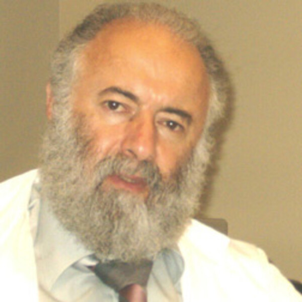
Alexander Velumian
PhD, DSc

Affiliations: Division of Neurosurgery, TWH-UHN
Courses Taught :
Co-Director, PSL444Y - Neuroscience 2 - Cellular & Molecular
Co-Director, JNR1444Y - Fundamentals of Neuroscience: Cellular and Molecular
Research Synopsis
Keywords: Spinal cord injury, axonal condunction, myelin, oligodendrocytes, glial-axonal interactions, patch clamp, imaging, spinal cord slices, in vitro, electrophysiology.
Detailed Description: My current research is focused on the physiology of myelinated axons and functional organization of living myelin sheaths in CNS white matter. We use a variety of rodent CNS white matter preparations for in vitro electrophysiology and imaging, such as spinal cord white matter, corpus callosum and optic nerves. Our approaches include compound action potential recording from white matter, whole cell patch clamp recording from oligodendrocytes, injection of fluorescent dyes into individual oligodendrocytes or myelin segments during whole cell recording or by microelectroporation, fluorescence imaging for visualization and 3-D reconstructions of dye-filled cytoplasmic networks of individual myelin sheaths. Our research is conducted in the spinal cord injury research program at the Toronto Western Hospital headed by Dr. Michael Fehlings.
My earlier work included intracellular microelectrode recordings combined with microiontophoretic administration of neurotransmitters on motoneurons using in vitro spinal cord preparations: (1973-1992: frog, immature rat, chick embryo; electrical/chemical transmission; ionic mechanisms of postsynaptic effects of glutamate, GABA and glycine, synaptic inhibition at early stages of development, myelination and the initial segment of the axon during the onset of myelination; whole cell patch clamp recordings: pyramidal neurons in hippocampal slices (1993-2000, combined with intracellular perfusion: effects of anions and intracellular Ca2+ buffering on various K channels and neuronal excitability), dissociated dorsal root ganglion neurons (2001-2002: HCN channels related to neuropathic pain).
METHODS USED
Cell and tissue culture: Brain slice, glial cells, hippocampal cells, neurons, oligodendrocytes.
Procedures: Fluorescence/confocal imaging, Infrared imaging, Sucrose gap recording, Brain slice, hippocampal cells, neurons, electrophysiology, in vitro electrophysiology, intracellular injection, patch clamp, voltage clamp
EQUIPMENT USED
Amplifier, digidata, electrophysiology rig, fluorescence microscope, fresh tissue sectioning systems, Micropipette puller, stimulator
PRESENT COLLABORATIONS
Outside the Department of Physiology:
Michael Fehlings, Surgery, U of T, Canada.
Recent Publications
Review:
Park E, Velumian AA, Fehlings MG. The role of excitotoxicity in secondary mechanisms of spinal cord injury: a review with an emphasis on the implications for white matter degeneration. J Neurotrauma 2004;21:754-774.
Velumian AA , Samoilova M. Chapter 1: White matter: Basic principles of axonal organization and function. In: Baltan S, Carmichael T, Matute C, Guochua X, Zhang JH, editors. White Matter Injury After Stroke & CNS Disorders. (United States): Springer; 2014. p. 3-38.
Appointments
Primary: Surgery.
