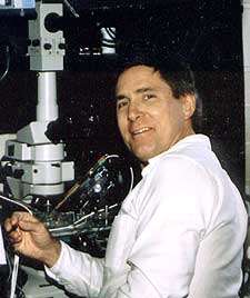
Milton Charlton, PhD
ACADEMIC STATUS
Departmental Status: Professor Emeritus
Primary/Cross-appointments: Physiology
Degrees: MSc 1971, PhD 1975
Courses taught: PSL 1026H, PSL374, JGY1555H
RESEARCH
Research Divisions: Brain Research and Integrated Neurophysiology B.R.A.I.N. Platform
Research Interests: Excitation-secretion coupling at synapses. Molecular organization of the presynaptic terminal. Mechanisms of neurotoxins. Confocal microscopy. Calcium signaling.
Keywords: Snap-25/ Syntaxin/ Synaptobrevin/ Snare/ Synapse/ Neuromuscular Junction Botulinum/ Presynaptic/ Botox/ Synaptic Vesicle/ Exocytosis.
Detailed Description:
We focus on the mechanism of transmitter release from presynaptic terminals. Our major strategy is to use pharmacological and biochemical interventions to deduce protein functions in exocytosis. For instance, injection of peptide fragments of specific proteins may interfere with the function of the native protein by making ineffective protein complexes. Injection of specific enzymes such as botulinum toxins which cleave proteins involved in exocytosis is another powerful technique. I like to use simple preparations which have presynaptic cells with distinct experimental advantages. For instance, much of my work used the giant synapse of the squid because here one can directly measure presynaptic membrane potential, presynaptic [Ca2+], presynaptic Ca2+ current and transmitter release while injecting proteins, peptides and other probes of protein function. At Toronto we use the crayfish neuromuscular junction because it has all the above advantages (except easy voltage clamping of presynaptic Ca2+ current) and, in addition, allows easy determination of spontaneous and evoked quantal transmitter release. Furthermore, crayfish synapses show a huge degree of synaptic differentiation; on one muscle fibre the probability of transmitter release is hundreds of times higher in synapses of phasic motorneurons than in synapses of tonic motorneurons. We hypothesize that there is a molecular differentiation which accounts for this difference. In this preparation we have been able to inject enzymes, peptides and antibodies into the presynaptic terminal while recording quantal transmitter release and presynaptic [Ca2+]. We also study the frog neuromuscular junction because it has a peculiar arrangement of the exocytosis machinery which allows easy visualization of protein distribution. Here, the active zones where transmitter exocytosis occurs are spaced at regular intervals of about 1 micron. This facilitates visualization of changes in vesicle and protein localization. Other preparations can be used when their particular advantages are required. For instance we are now working on exocytosis from nociceptors.The laboratory contains setups for intracellular injection and recording, voltage clamping, confocal imaging, and wide field fluorescence imaging.
METHODS USED
Cell and tissue cultures: Brain cell.
Procedures: Electrophysiology, immunocytochemistry, intracellular injection, intracellular membrane potential, ion imaging using widefield and confocal microscopy, neuromuscular junction, path clamp, proteomics, synaptic potential, voltage clamp, western blot.
EQUIPMENT USED
Amplifier (Gradient PCR cycler), confocal microscope, culture hood (Sterile class 2), culture incubators, electrometer, EMCCD, fluorescence microscope (Upright, Widefield), intracellular sharp electrode measurement of membrane potential, monochromator (Rapid switching, for Fura2 ration ion imaging/ with EMCC camera), motorized micromanipulators, simultaneous patchclamp and Fura2 imaging, 2D gels.
SAMPLE PUBLICATIONS

*Dr. Charlton has retired and is not taking on new students
Phone: (416) 978-6355
Fax: 416-978-4940
Email: milton.charlton@utoronto.ca
Address: Department of Physiology
Medical Sciences Building
1 King's College Circle
University of Toronto
Toronto, ON, M5S 1A8
